About
Mission Statement
The mission of Thai Microcosmos is to promote microscopy exploration and microscopy education throughout Thailand. We achieve this by sharing discoveries of the invisible world around us, and offering opportunities for public engagement and education about the fascinating microscopic life that exists in our everyday environment.
Microscopy reflects a confluence of a number of my interests and hobbies. I have always held a strong interest in science, from astronomy and astrophotography to physics and biology. For more than 20 years I have married my passion for the night sky with my interests in photography and digital processing, and during that entire time I imagined turning my camera inward toward the microcosmos.
In 2022, during a visit home to the USA, I was gifted an amazing microscope – a Nikon Inverted Diaphot with Phase Contrast (circa 1985). Weighing more than 65 pounds, it was not a simple task bringing this scope with me from the USA to my home in Bangkok, but it made it in one piece. I have the following objectives:
- Nikon 4/0.20 Plan APO
- Nikon 10/0.25 Plan Phase Contrast
- Zeiss 16/0.40 NeoFluor PH2
- Nikon 20/0.75 Plan APO
- Nikon 40/0.65 Plan Phase Contrast
- Nikon 40/1.30 Fluor Oil Immersion
- Nikon 40/1.0 Plan APO Oil Immersion
- Nikon 100/1.30 Fluor Oil Immersion

The microscope came with Nikon’s ELWD (Extra Long Working Distance) 0.30 N.A. condenser, which served me well for the first year. However I later changed to Nikon’s LWD 0.52 N.A. condenser, which performs better and allows me to achieve dark field using the Zeiss 16/0.40 objective.

The microscope is binocular, and also has dedicated camera and videocam ports. The Nikon F Lens mount allows my Nikon D750 DSLR to attach and reach focus without an adapter. HDMI out from the camera allows live views on our TV or a computer monitor. The camera records images at 24 megapixels, and video at 1080P.
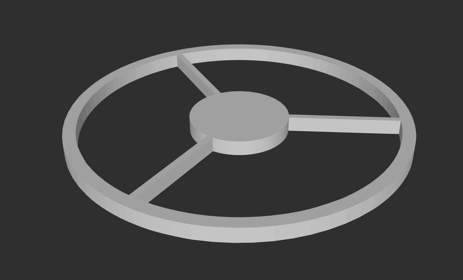
I began experimenting in 2023 with homemade dark field and rheinberg filters. Hand cut black paper and blue cellophane were taped to clear 45mm watch glass crystals, and while this worked the filters lacked the performance that perfect circles would achieve. In May, 2024 I designed my own set of 3-d modeled dark field patch stops and had them printed for me. The results are a noticeable improvement.

In addition to Phase contrast, during my 2023 trip home I was given a complete Nikon Fluorescence System with Xenon light sourcefor the microscope. I’ve begun playing with various dyes, including Acridine Orange, Mitotracker Green and Mitotracker Red.

Physical Modifications
The microscope’s internal AC power source powers a 50W halogen bulb. There is a dial that allows switching from 110V to 220V, but I didn’t want to trust it and risk frying the electronics, so instead I began by using a step down transformer. However, initial videos showed screen flicker unless the camera shutter speed was set to a multiple of 50 (AC power in Thailand is 50hz), and this became quite limiting when I wanted slower shutter speeds to allow more light. With the help of a friend, I modified the scope to bypass the internal power source and instead use an external 12V 6A DC laptop-style DC power brick. We included rheostatic control using a 13 Mhz PWM, and the result was that I could shoot at any shutter speed flicker-free.

Update: After 1-1/2 years of use, the PWM board failed. As it turns out, while sold as a 40A PWM, it was really a 10A PWM, and various components melted over time. I have since replaced it with a true 40A PWM controller in its own box. While this new controller doesn’t have a feature to bypass the rheostatic control, I can quickly bypass the PWM box using the quick-release power connecters that we used to operate the lamp at 100% for purposes of burning off built-up residue.

Next, my wife’s cousin designed and 3D printed a replacement center disk for the stage, with a groove and magnetic top to hold slides in place. This is critical for use with the oil immersion objective. When using an inverted microscope the slide is placed with the coverslip facing down. The objective is brought into place from underneath, but once an oil immersion lens contacts the coverslip any additional pressure merely pushes the slide up off the stage surface, preventing the spring-loaded objective tip from focusing deeper into the slide. The magnetic top solves this issue, applying counter pressure to the slide and allowing the oil immersion objective to focus further into the sample.
In November 2023 I switched to a 100W halogen light source, running it off a more powerful transformer. The additional light allows video capture at much lower ISO, greatly reducing digital noise.
I’ve given all optical surfaces (both the microscope and camera) that I can reach a thorough cleaning. I’ve also become confident enough to open up the scope, remove the prisms and mirrors and clean them. This elimated stubborn dust spots that I lived with for almost a year. I now regularly clean all optical surfaces as well as the camera sensor every 6-8 weeks, as I’m in a quite dusty environment. I had a plastic cover made for the scope which is kept on whenever the scope isn’t in use.

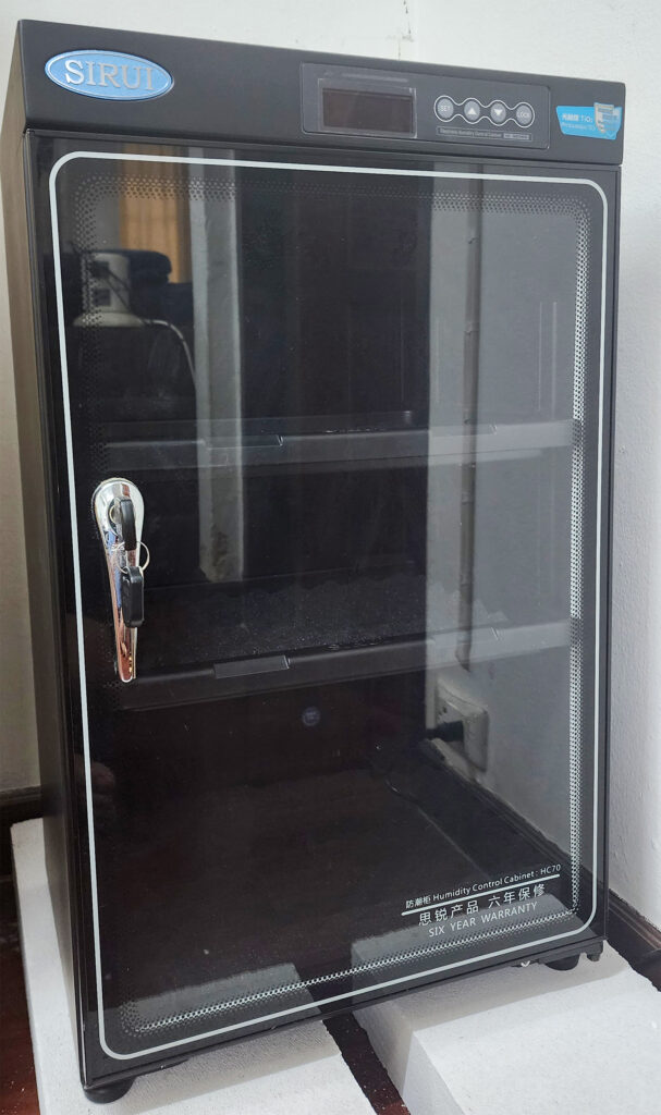
I use a Sirui HC70 70-liter humidity control cabinet to store optical components while not in use. The humidity level is set to 42%.

Future enhancements include plans to develop an improved stage, one that will have joystick control to allow smoother movement (especially diagonally) while following living organisms, as well as location memory to mark items of interest that can easily be returned to at the click of a mouse.
My workspace:


Advancing the Hobby
In early 2023 I started a 20L pondwater microorganism aquarium to provide a constant stream of organisms to view. I periodically refresh the aquarium with more water and plants from the original source (a fishing pond 30 minutes away). In November, 2023 I began playing around with microscopic crystal formation using various chemicals around the house.

Public Education
Using cables and other equipment I already had, I am able to live stream the microscope on YouTube. In line with the many years of sidewalk astronomy and public education I participated in, I am currently having conversations with several Bangkok primary and secondary schools to offer complimentary “online field trips” to science classes, offering views they might never get using equipment in the classroom (if they even have access to microscopes and live samples).


